Our commitment to quality care starts with helping patients learn more about the exams we provide, including what to expect during an exam. We provide a broad range of ultrasound services, including:

Abdominal ultrasound
Ultrasound imaging evaluates the liver, pancreas, gallbladder, bile ducts, spleen and kidneys. Referring providers order abdominal ultrasound imaging for a number of reasons. The most common reasons are abdominal pain, elevated lab values, nausea, vomiting or diarrhea.
Abdominal ultrasound FAQ
- What preparation is required? Patients are required to have nothing to eat or drink after midnight the day before their ultrasound exam.
- Can I still take my medications before the exam? You may take your medications before your exam; just be sure you take them only with water.
- Can ultrasound evaluate for stomach ulcers? No, ultrasound is not the best imaging mode to evaluate the stomach or bowel because both contain air. Air and ultrasound are not diagnostically compatible. Ultrasound waves are unable to move through air diagnostically, this makes ultrasound evaluation of the stomach or bowel very limited.
- Who interprets the abdominal exam? Sound Health Imaging contracts with Proscan Reading Services to read our exams. They are fellowship-trained, board-certified radiologists with subspecialty training.
- When and how will I get results? The abdominal exam will be read by the radiologist within 24 hours of your study. The results will be sent directly to your referring provider the weekday after your exam.
- How do I schedule my abdominal ultrasound at Sound Health Imaging? A provider order is required for the abdominal ultrasound. When you see your health care provider, let them know you would like to have your ultrasound scheduled at Sound Health Imaging. Or, after you see your provider, you can call our office and make your ultrasound appointment and then bring your order with you at the time of exam.
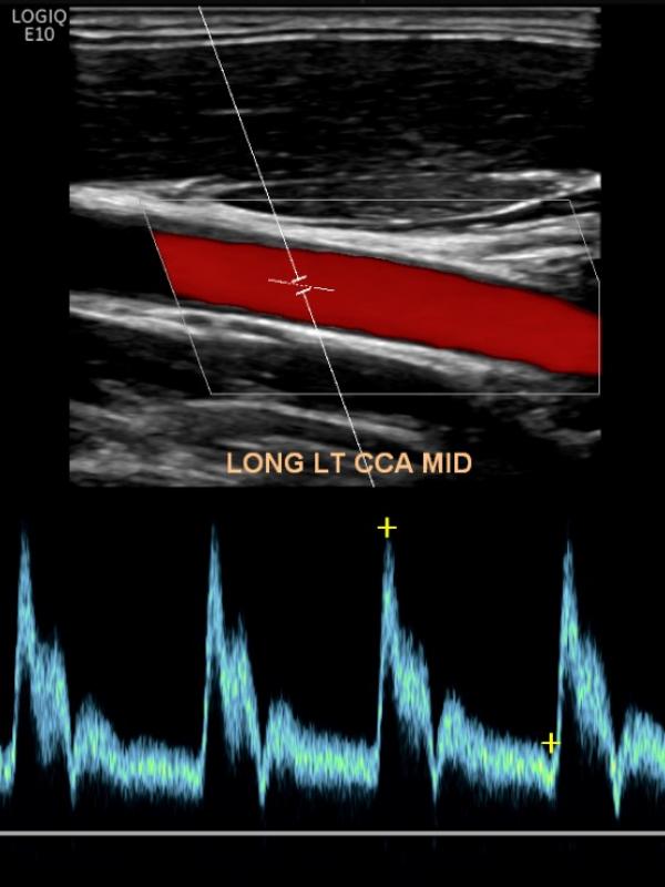
Carotid ultrasound
Ultrasound imaging of the carotid arteries evaluates for plaque build-up or blockage. Referring providers order carotid ultrasound for a number of reasons. The most common reasons are: dizziness, stroke, blurred vision, or bruit discovered on physical exam.
Carotid ultrasound FAQ
- Is there any required preparation for a Carotid Ultrasound? No preparation is required.
- Who interprets the Carotid ultrasound exam? Sound Health Imaging contracts with Proscan Reading Services to read our exams. They are fellowship-trained, board-certified radiologists with subspecialty training.
- When and how will I get results? The Carotid ultrasound exam will be read by the radiologist within 24 hours of your study. The results will be sent directly to your referring provider the week-day after your exam.
- How do I schedule my Carotid ultrasound at Sound Health Imaging? A provider’s order is required for the Carotid ultrasound. When you see your healthcare provider, let them know you would like to have your ultrasound scheduled at Sound Health Imaging. Or, after you see your provider, you can call our office and make your ultrasound appointment and then bring your order with you at the time of exam.
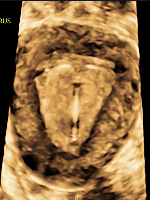
Female Pelvic/Gynecologic ultrasound
A provider may order a pelvic ultrasound for a number of reasons. Some common reasons are: pelvic pain, irregular bleeding, lack of periods or fertility assessment. Ultrasound has proven to be an important diagnostic tool in evaluating the gynecologic patient. With improved technology, ultrasound plays an integral role in evaluating the female pelvis. The uterus, uterine lining (endometrium), and ovaries are well-visualized using transabdominal and transvaginal ultrasound imaging.
Female pelvic/gynecologic ultrasound FAQ
- What should I expect during this exam? Typically, your provider will order a transabdominal and transvaginal ultrasound to evaluate the uterus, uterine lining and ovaries. Transabdominal (ultrasound performed by placing the transducer on the skin of the lower abdomen). Transvaginal (ultrasound performed by placing a cylinder-shaped ultrasound camera into the vagina). Transvaginal ultrasound adds information and is often the preferred method of evaluation.
- What is the preparation for a Pelvic ultrasound? Patients are asked to come to the exam with a full bladder. We recommend drinking 24 ounces, finishing 45 minutes before your appointment. You can drink any kind of clear liquid, including pop, tea, juice or water. No milk products, as they do not fill the bladder.
- Who interprets the pelvic exam? Sound Health Imaging contracts with Proscan Reading Services to read our exams. They are fellowship-trained, board-certified radiologists with subspecialty training.
- When and how will I get results? The pelvic exam will be read by the radiologist within 24 hours of your study. The results will be sent directly to your referring provider the week-day after your exam.
- How do I schedule my Pelvic ultrasound at Sound Health Imaging? A provider order is required for a pelvic ultrasound. When you see your healthcare provider, let them know you would like to have your ultrasound scheduled at Sound Health Imaging. Or, after you see your provider, you can call our office and make your ultrasound appointment and then bring your order with you at the time of exam.
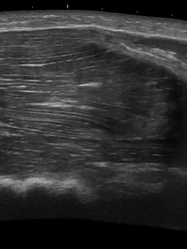
Musculoskeletal ultrasound (MSK)
Ultrasound imaging of tendons, ligaments, and muscle groups. Referring providers order musculoskeletal ultrasound for a number of reasons. Some common reasons are: suspicion of muscle or ligament/tendon tear or damage, trigger finger or lump.
Musculoskeletal ultrasound FAQ
- What is the preparation needed for my MSK ultrasound? No preparation is necessary.
- Who interprets the MSK ultrasound? Sound Health Imaging contracts with Proscan reading services and Specialist Direct reading services. They are fellowship-trained, board certified radiologist with subspecialty training.
- When and how will I get results? The MSK ultrasound exam will be read by the radiologist within 24 hours of your study (unless the study indicates need for results sooner). Final results will be sent directly to your referring provider the week-day after your exam.

Obstetric ultrasound
All sonographers at Sound Health Imaging are nationally board-certified in Obstetrics and Gynecology (Ob/Gyn) ultrasound by the American Registry of Diagnostic Sonographers.
Ultrasound Timeline in Pregnancy
- Early 1st Trimester Ultrasound: In the first trimester, ultrasound exams are done to demonstrate the viability of the pregnancy and confirm a due date. A heartbeat can be detected at about 6 weeks menstrual age. The 1st trimester is the most accurate time to date the pregnancy.
- 11-13 Week Genetic Screen (Ultrascreen): Screens for the likelihood of chromosomal abnormalities, specifically Down’s syndrome (Trisomy 21), Trisomy 13, and 18. Consists of a measurement of the nuchal translucency (a space behind the fetal neck) and a finger poke for blood droplets. Detection rates for chromosomal anomalies are 91-95% with experienced sonographers performing the study.
- Fetal Anatomical Survey (best results at 20 weeks): This is a comprehensive ultrasound that evaluates the entire fetal anatomy, fetal size, placenta, and maternal uterus, cervix and ovaries. Fetal Anatomy that is imaged in detail is the fetal: brain, face, spine, arms, legs, hands, feet, stomach, kidneys, bladder and umbilical cord. The exam will also focus a large portion of time on viewing the fetal heart in detail. Virtually all congenital heart defects are detected now, with the heartbeat heard and all four chambers of the heart seen. It is now also possible to see the aorta rising from the left ventricle and the pulmonary artery from the right, as well as the returning veins into the right and left aorta with its branches. Finally, this is the time when fetal gender is easily determined for parents who would like to know.
- Third Trimester Ultrasounds: Ultrasound examinations in the third trimester are ordered on a case-by-case basis and depend on the referring provider’s monitoring requirements of mother or baby.
- 3D/4D Ultrasounds: The best time in the pregnancy for 3D/4D imaging is between 26 and 30 weeks gestational age. At this time there is plenty of amniotic fluid around the baby. Fluid enhances the images and adequate amniotic fluid around the baby is essential for good 3D/4D imaging. As the baby gets closer to term the amniotic fluid naturally decreases, making it more difficult to get good 3D/4D images. Also, 26-30 weeks is a good time for the 3D/4D because the baby has started to “fill out” and take on some of the features it will have at birth. Before 26-30 weeks gestational age the baby looks more skeleton-like. There are a number of factors that affect the quality of the 3D/4D images. Ideally, the baby is positioned away from the placenta and uterine wall. If the fetal hands, feet or umbilical cord are in front of the face it will result in less than ideal images. In order to have a 3D/4D ultrasound at Sound Health Imaging you must have a referral from your medical provider. During the 3D/4D ultrasound, measurements will be taken to assess fetal growth, but an anatomical survey of the baby will not be done. We require that a previous 20-22 week ultrasound for fetal anatomical surveillance has already been done prior to the 3D/4D ultrasound. 3D/4D ultrasounds are not medically necessary and cannot be billed out to insurance companies. Please contact our office for more information.
Obstetric ultrasound FAQ
- What is the preparation for my Obstetric ultrasound? We ask 1st and 2nd Trimester patients come in with a full bladder. We recommend drinking 24 ounces, finishing 45 minutes before your appointment. You can drink any kind of clear liquid, including pop, tea, juice or water. No milk products, as they do not fill the bladder. No preparation is needed for patients who are in the 3rd Trimester.
- Who can come into the ultrasound exam with me? We allow anyone in the exam room the mom wishes to invite.
- Can we bring children to the ultrasound appointment? Children are welcome if another adult is along to provide supervision. The 20-week fetal survey scan takes approximately 45-60 minutes to complete, and small children tend to get bored quickly.
- When can I find out the baby’s gender? During the 20 week, fetal anatomy scan the sonographer is happy to tell you the gender if parents wish to know!
- Who interprets my obstetric ultrasound? Our reading physicians are comprised of highly specialized, board certified radiologists and maternal fetal medicine specialists.
- When and how will I get results? The obstetric ultrasound will be read by the physician within 24 hours of your study. The results will be sent directly to your referring provider the week-day after your exam.
- How do I schedule my Obstetric ultrasound at Sound Health Imaging? A provider order is required for the obstetric ultrasound. When you see your healthcare provider, let them know you would like to have your ultrasound scheduled at Sound Health Imaging. Or, after you see your provider, you can call our office and make your ultrasound appointment and then bring your order with you at the time of exam.
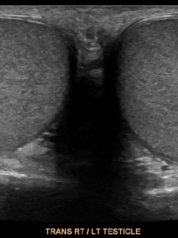
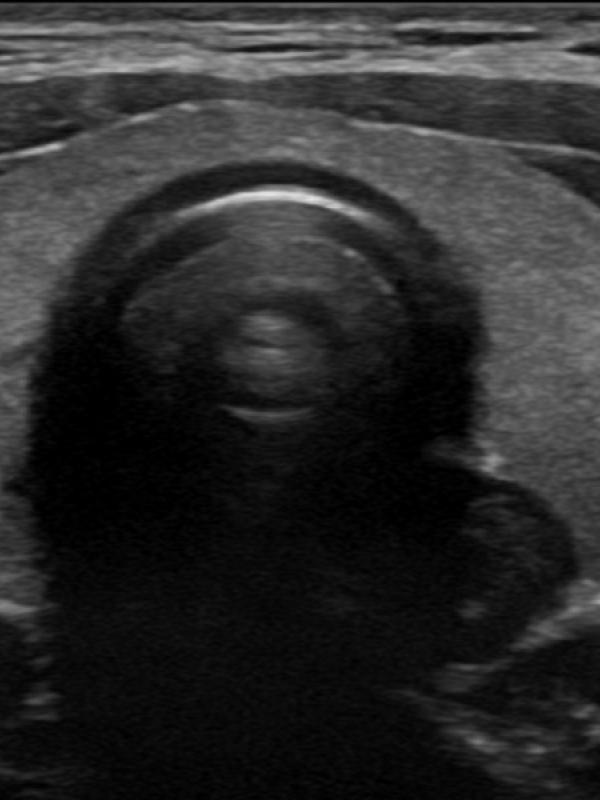
Scrotal ultrasound
Ultrasound imaging is the primary diagnostic modality used to evaluate the scrotum/testicles. Referring providers will order a scrotal ultrasound for a number of reasons. The most common reasons are: testicular pain, a lump is discovered, or if a hernia is suspected. Undescended testis.
Scrotal ultrasound FAQ
- Will the ultrasound of the scrotum and testicles be painful? No. The ultrasound exam will not be painful at all. The sonographer uses warm ultrasound gel and very light pressure from the transducer to image the testicles and scrotal contents.
- Is there any preparation required for a scrotal ultrasound exam? No, there is no preparation needed before having a scrotal ultrasound.
- Who interprets the scrotal exam? Sound Health Imaging contracts with Proscan Reading Services to read our exams. They are fellowship-trained, board-certified radiologists with subspecialty training.
- When and how will I get results? The scrotal ultrasound exam will be read by the radiologist within 24 hours of your study unless the study indicates need for results sooner. The results will be sent directly to your referring provider the week-day after your exam.
- How do I schedule my Scrotal ultrasound at Sound Health Imaging? A provider order is required for the scrotal ultrasound. When you see your health care provider, let them know you would like to have your ultrasound scheduled at Sound Health Imaging. Or, after you see your provider, you can call our office and make your ultrasound appointment and then bring your order with you at the time of exam.
Thyroid ultrasound
Ultrasound an excellent diagnostic tool for thyroid evaluation. It is used to evaluate the size of the gland, texture, and evaluate for lesions. The most common reasons for a referring provider to order a thyroid ultrasound are for abnormal thyroid lab values or a suspected abnormality following a physical.
Thyroid ultrasound FAQ
- Is there any required preparation for a Thyroid ultrasound? No, there is no prep for a thyroid ultrasound.
- Who interprets the Thyroid ultrasound exam? Sound Health Imaging contracts with Proscan Reading Services to read our exams. They are fellowship-trained, board-certified radiologists with subspecialty training.
- When and how will I get results? The Thyroid ultrasound exam will be read by the radiologist within 24 hours of your study. The results will be sent directly to your referring provider the week-day after your exam.
- How do I schedule my Thyroid ultrasound at Sound Health Imaging? A provider order is required for the Thyroid ultrasound. When you see your health care provider, let them know you would like to have your ultrasound scheduled at Sound Health Imaging. Or, after you see your provider, you can call our office and make your ultrasound appointment and then bring your order with you at the time of exam.

Venous ultrasound
Ultrasound imaging of the venous system is used to evaluate the vessel and returning blood flow from upper or lower extremities. Referring providers order venous ultrasounds for a number of reasons. The most common reasons are: leg pain or swelling, varicose veins, or evaluation for a suspected blood clot.
Venous ultrasound FAQ
- What is the preparation for a venous ultrasound examination? There is no required preparation for a venous ultrasound.
- What is a vein mapping ultrasound? A detailed evaluation of the entire venous system in your lower extremities. The sonographer will first evaluate for a blood clot, then will map out the superficial venous system and evaluate for valve incompetency
- Who interprets the Venous ultrasound exam? Sound Health Imaging contracts with Proscan Reading Services to read our exams. They are fellowship-trained, board-certified radiologists with subspecialty training. In some instances, with venous mapping exams, the referring provider will read the study.
- When and how will I get results? The Venous ultrasound exam will be read by the radiologist within 24 hours of your study unless the study indicates need for results sooner, or if the referring physician has requested results immediately following the exam. The results will be sent directly to your referring provider the week-day after your exam.
- How do I schedule my Venous ultrasound at Sound Health Imaging? A provider order is required for the Venous ultrasound. When you see your health care provider, let them know you would like to have your ultrasound scheduled at Sound Health Imaging. Or, after you see your physician, you can call our office and make your ultrasound appointment and then bring your provider order with you at the time of exam.